Epilepsy surgery
Epilepsy surgery is a treatment approach for some people with epilepsy whose seizures are not controlled by medication. By removing part of the brain where seizures begin, seizures can occur less often, or not at all. Surgery can be considered when seizures begin from a small part of the brain (‘focal epilepsy’). Only parts of the brain that can be removed safely are considered for surgery so that essential brain functions continue to work. Detailed testing and assessment is usually required before someone can be offered epilepsy surgery.
There are several different types of epilepsy surgery. The most common is temporal lobectomy, where part of the temporal lobe of the brain is removed. Other procedures include lesionectomy – removal of a brain lesion that is causing seizures or corpus callosotomy – cutting the corpus callosum to prevent seizures spreading from one side of the brain to the other.
New technologies are being used to treat even smaller brain regions. These include radiofrequency thermocoagulation (RFTC) where a wire is placed inside the brain and electric current creates a small lesion around the tip. MRI-guided high-frequency focused ultrasound (HIFU) is a new minimally-invasive approach being researched. Here ultrasound waves are applied from outside the head to remove a precisely targeted lesion deep within the brain.
How The Florey is making a difference
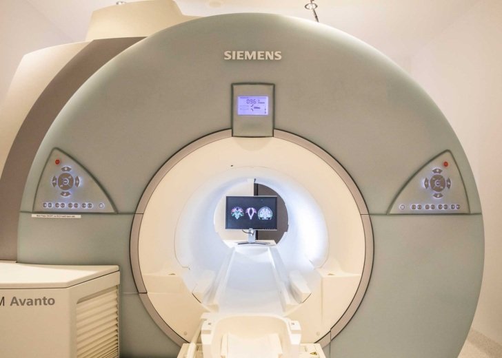
At the Florey we’re developing advanced MRI techniques to assist in epilepsy surgery planning, including:
- functional MRI techniques that use in-scanner EEG to identify the origin of seizures in the brain
- diffusion imaging techniques that allow us to map connections between brain regions and identify how epilepsy changes brain networks
- artificial intelligence methods to identify brain abnormalities before and after epilepsy surgery.
Identifying the brain region where seizures begin is the first challenge for making epilepsy surgery available. Through the Australian Epilepsy Project, the Florey is researching improvements for MRI scans of the brain, to better detect brain lesions or small abnormalities in brain shape. This project has already changed the lives of people living with epilepsy, by revealing a brain lesion that had not been previously detected.

When brain structure appears normal, figuring out where seizures are starting from is even more challenging. This is the goal of the Florey AFFECT EEG-fMRI study, which uses functional MRI scanning at the same time at brain-wave recording (EEG), to pinpoint where in the brain epilepsy activity is occurring. This ongoing clinical trial will evaluate the clinical impact of early access to this information.
More information
While The Florey researches epilepsy, the institute does not offer a clinical or diagnostic service and we are unable to give advice on treatment in individual cases or provide referral advice. You can find more information about epilepsy surgery at the following links.