Research highlights
- Multiple sclerosis reduces the number of nerve fibres both in affected nerves but also in seemingly unaffected nerves
- Advanced magnetic resonance imaging (MRI) can now identify subtle damage to connections within the brain, helping neurologists monitor and treat patients
- MS patients with a history of vision loss had reduced nerve fibre density in the pathways taking visual information from the eyes to the rest of the brain
- The new FBA techniques will allow researchers to fast track early stage clinical trials, monitor patients’ response to new therapy and predict disease progression.
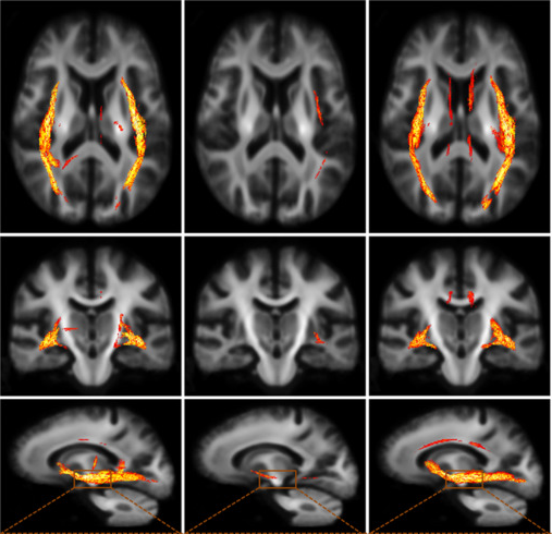
Scientists from the Florey Institute of Neuroscience and Mental Health and the University of Melbourne have used a new MRI technique to examine how multiple sclerosis (MS) affects the visual nerve pathway in patients.
Many people living with MS experience inflammation of their optic nerves, causing dim and blurry vision, eye pain and sometimes temporary blindness. This is obviously very distressing for patients and their families, but the condition usually resolves within a couple of weeks. What damage the inflammation does to other parts of the brain’s visual pathways however is not well known.
The new imaging technique, developed at the Florey, can potentially allow researchers to reduce the size and duration of early stage clinical trials, monitor patients’ progress on therapy and predict how their disease will progress.
Currently, standard MRI in the hospital setting only allows clinicians to see MS lesions in the brain. The Florey’s new imaging analysis technique, developed by Professor Alan Connelly, now allows researchers to see microscopic damage to the brain that previous MRI techniques missed.
“This new method allows us to identify reductions of nerve fibres connecting different regions of the brain. We can detect and characterise damage to brain pathways to a degree that no-one has been able to achieve previously.”
In a patient with multiple sclerosis, the protective fatty sheath surrounding nerve fibres is attacked by the body’s own immune cells. This leaves the nerves vulnerable to attack by those same cells. The authors applied their analysis method to study people with MS who previously experienced vision issues and found a very specific loss of the density and diameter of nerve bundles that conduct visual information from the eye to the brain.
Dr Scott Kolbe, who led the current research that was published in NeuroImage: Clincal, says, “Using this new MRI technique, we are seeing for the first time how the connections in the brain degenerate in people with MS. This may allow us to better select patients for clinical trials, monitor disease progression and to choose certain patients for specific therapies.” Importantly, the new imaging technique does not require any new hardware, allowing existing hospital MRI scanners to acquire suitable images from patients to enable the new connectivity maps to be generated, creating the potential for treating clinicians to optimise their treatment for individual patient needs.
Dr Kolbe’s team are doing further research to look at the links between nerve fibre damage and changes in coordination during walking, writing and even speaking in people with MS. They hope to uncover how damage to specific parts of the brain leads to the variety of symptoms suffered by people with MS.
Scientists are still unsure of the exact mechanisms causing multiple sclerosis, but it tends to occur more commonly in certain populations, one of which is young women. Dr Darshini Ayton is a research fellow and lecturer at Monash University. Darshini was diagnosed with the most common form (‘relapsing-remitting’) of MS in 2008.
“Current MRI imaging doesn’t always provide the full picture. There have been times when I have had MS symptoms but there have been no changes in my MRI and other times when I have had MRI changes but experienced no symptoms. As a patient this can be frustrating and confusing. I am hopeful that this new MRI technique will provide better information both for clinicians and patients in relation to prognosis and treatment impacts.”
The team are currently looking for healthy volunteers to further improve the MRI analysis technique. If you are interested in volunteering, please contact [email protected]