Neurohistology Laboratory
About us
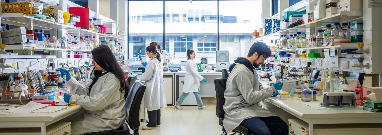
The Florey’s Neurohistology Laboratory provides specialised histology services for preparing and examining both diagnostic and research tissues for the Institute’s research staff, students and external clients on a cost-recovery basis.
The Neurohistology Laboratory also provides services to many medical diagnostic groups including the Victorian Brain Bank, the Australian National CJD Registry, the Royal Melbourne, the Alfred, St Vincent’s and the Royal Hobart hospitals, and South Australia pathology.
Highly qualified staff with a broad knowledge of human and animal histopathology are available to provide expertise and services to assist in the analysis of changes observed in tissue microarchitecture present in a variety of neurodegenerative diseases and research tissues.
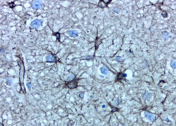
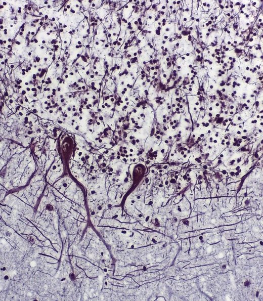
Image top-right shows brain astrocytes labelled with an antibody to glial fibrillary acidic protein. Magnification 800x. Image courtesy of Dr Ian Birchall.
Services
Services in the laboratory include:
- Preparing and processing human and experimental animal tissues for paraffin embedding.
- Serial and series sectioning (2 – 8 micron) of paraffin-embedded tissues.
- Chemical staining techniques – H&E, Masson’s trichrome, PAS and others.
- Immunohistochemical labeling techniques with antibodies and lectins.
- Microtome training and hire for client use.
- Immunohistochemical method development for client supplied antibodies.
- Macroscopic and microscopic digital imaging.
Neurohistology staff are available to provide project consultation and itemised quotes for studies requiring histological preparations and assessments.
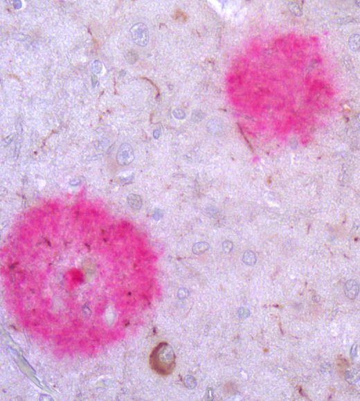
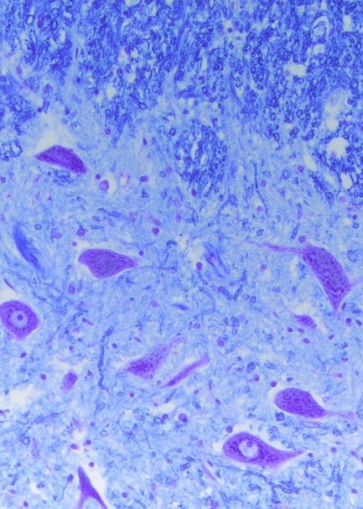
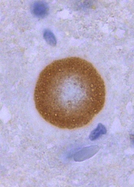
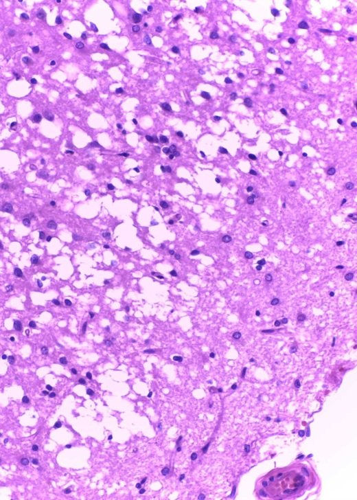
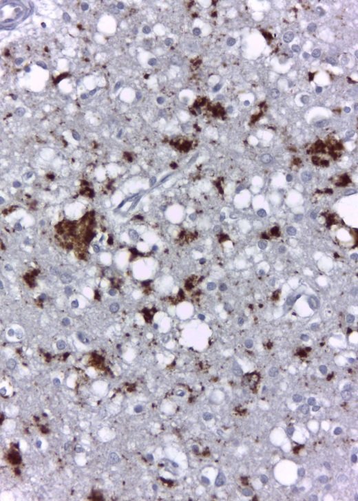
Histological images
Explore some of the images produced within the facility.
Contact
Neurohistology Laboratory
The Florey
Level 4, Kenneth Myer Building
30 Royal Parade
Parkville Victoria 3010
Facility staff
Enie Lei