Neuropix
Illuminating the wonders of the brain
Research at The Florey is geared at unravelling the complexities of neural circuits, studying neurodevelopmental processes, investigating disease mechanisms, and exploring potential therapeutic strategies for neurological disorders.
Microscopy techniques play a critical role in advancing our understanding of the brain’s structure and function. The Neuropix Imaging competition highlights these research efforts through a microscopic lens.
We hope to inspire the next generation of researchers through the power of imaging.
2026 winners
Fourth of July
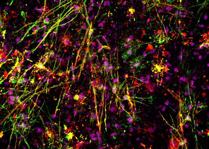
Scientific Award – Francois Beau, PhD student, Myelin in Health and Disease Group
This is an image of a lab-grown human mini-brain, in which we can see oligodendrocyte cell bodies (purple) as well as their projections (green and red).
The First Spark
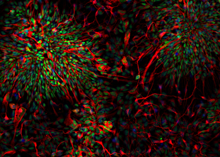
People’s Choice Award – Gabriella Farrell, PhD student, Stem Cells and Neural Repair Group
Using stem cells grown in the laboratory, this image recreates one of the earliest stages of human cortical development. The cells spontaneously organise into neural rosettes, revealing the remarkable capacity of human stem cells to build the earliest blueprint of the developing cortex.
2026 entrants
Microglial Convergence
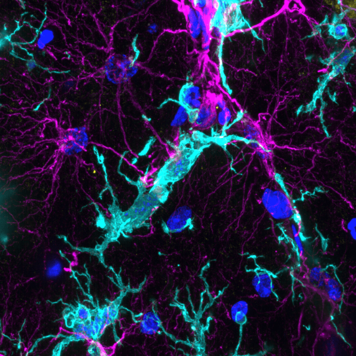
Angela Connelly, Research Assistant
Microglia (teal) and astrocytes (magenta) independently detect injury-associated signals and converge at sites of tissue damage and neuroinflammation in the sheep hippocampus. Nuclei are shown in blue.
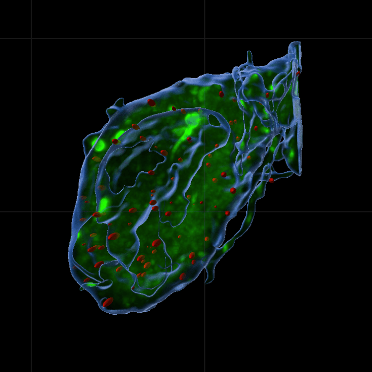
Varicella Neuron
Dr Chiara Pavan, Research Fellow
This is a neuron derived from a patient with Parkinson’s disease and how it looks in his disease stage, with pathological granules highlighted in red.
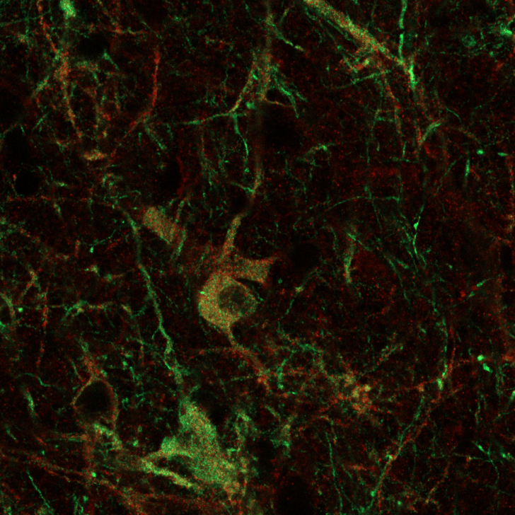
Neuronal Forest
Dr Chiara Pavan, Research Fellow
This is a stem cell derived neuronal graft in a rodent brain used to study Parkinson’s disease. Dopamine neurons are labelled in red and green.
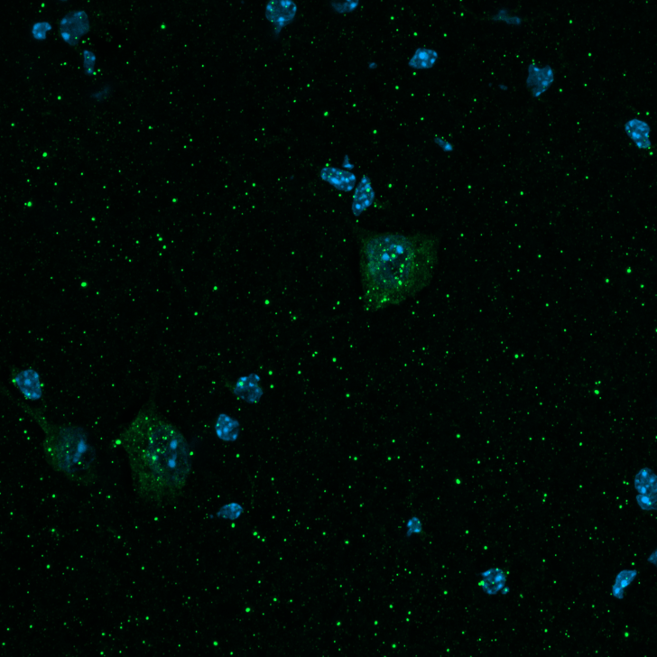
Intercellar
Emily Fraser, Master’s Student
This image shows neurons (labelled in green) that have been transplanted into a mouse brain, and the nuclei of cells are shown in blue.
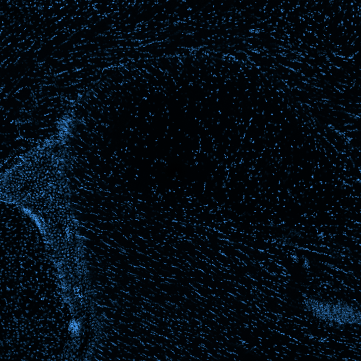
The Great Nuclear Tidal Wave
Emily Fraser, Master’s Student
This image shows a blue stain revealing the nuclei of mouse cells. The natural arrangement creates a striking wave-like pattern throughout the brain tissue.
Bioluminescent Bay
Emily Hadzajlic, PhD student
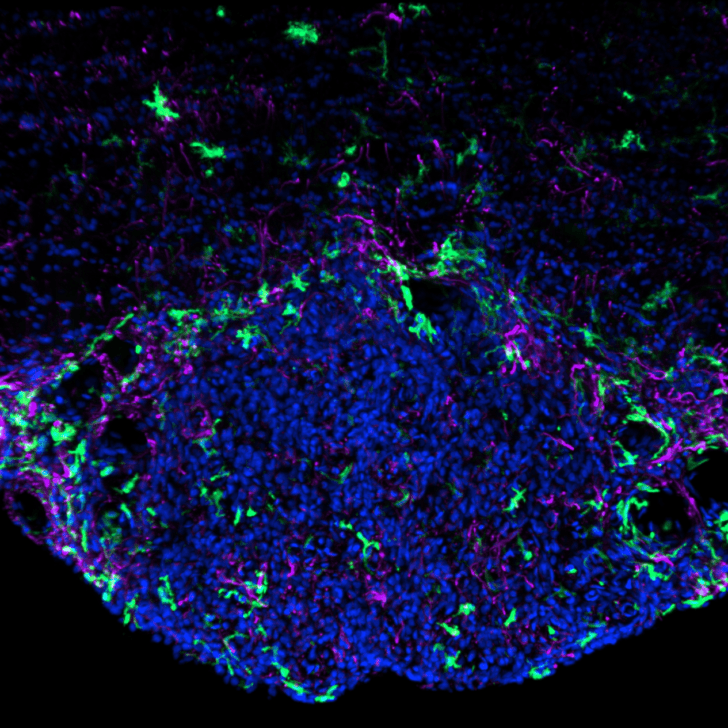
This image shows microglia (labelled in teal), astrocytes (labelled in purple) and nuclei (labelled in blue) in the subfornical nucleus of the rat brain. The subfornical nucleus is densely packed, as can be seen. It is one of the rare brain regions lacking the protective blood brain barrier, exposing it directly to anything flowing through the blood. This image reminds me of the bioluminescent algae sometimes seen glowing at night on the ocean. Like waves breaking on the shore, signals from the blood inundate the subfornical nucleus, where they are processed before being transmitted to other brain regions.
Constellations
Gabriella Farrell, PhD student
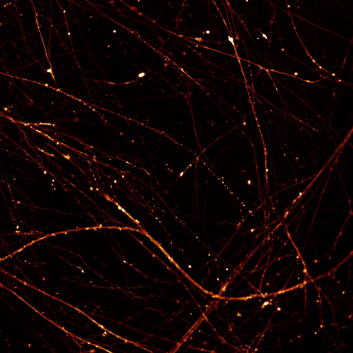
This image shows a network of mature human brain cells grown from stem cells in the laboratory. Their long, branching extensions form an intricate web of connections, resembling constellations scattered across the night sky. Visualised using a fluorescent dye that responds to calcium, these extensions provide the pathways through which brain cells communicate.
Woven Together
Gabriella Farrell, PhD student
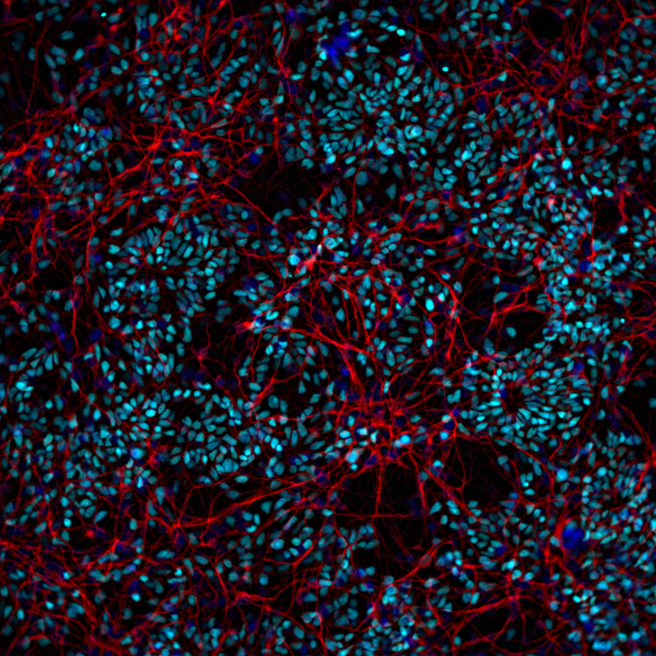
This image recreates an early stage of human cortical development using stem cells grown in the laboratory. As the cells organise themselves, newly formed neurons extend long, branching connections around clusters of developing brain cells, creating an intricate network. Fluorescent markers reveal this remarkable process of self-organisation, illustrating how the foundations of the cortex begin to take shape.
Neurogenesis in Bloom
Montanna Waters, PhD student
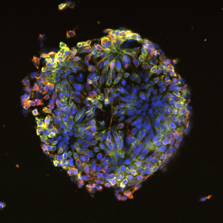
This image captures stem cells in the process of transforming into early brain cells, forming a structure known as a neural rosette. Grown on a specially designed surface, the cells arrange themselves into swirling, blooming patterns that resemble the structures seen in a developing embryo’s brain. Colour reveals different aspects of this process: green marks a protein that helps the cells stick together as they organise, red highlights cells on their way to becoming neurons, and blue marks each individual cell’s nucleus.
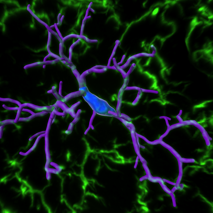
Ewe-nique Microglia
Sarah Brooke, PhD student
Microglia 3D remodelling. Sample imaged from brain tissue collected from a merino ewe (female sheep). This image shows microglia (Iba1 staining labelled in green), with the 3D model representing the cell soma (blue) and branches (purple).
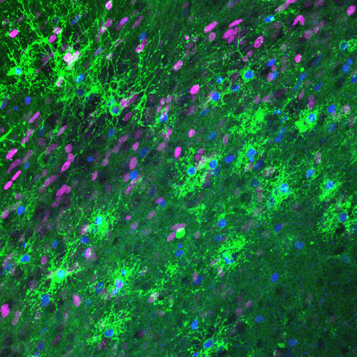
Paint Splatter Brain
Victoria Lee, Honours student
Pictured here are oligodendrocytes (cells that produce myelin, the protective layer surrounding brain axons) labelled in green, with their nuclei shown in blue. Magenta denotes proliferating cells.
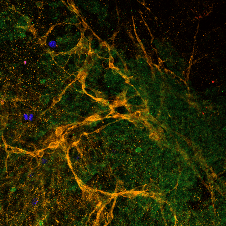
Caught in an Oligo Web
Victoria Lee, Honours student
Pictured here are immature oligodendrocytes (cells that produce myelin, the protective layer surrounding brain axons) in orange, branching out between cell nuclei stained green. Blue denotes proliferating cells.
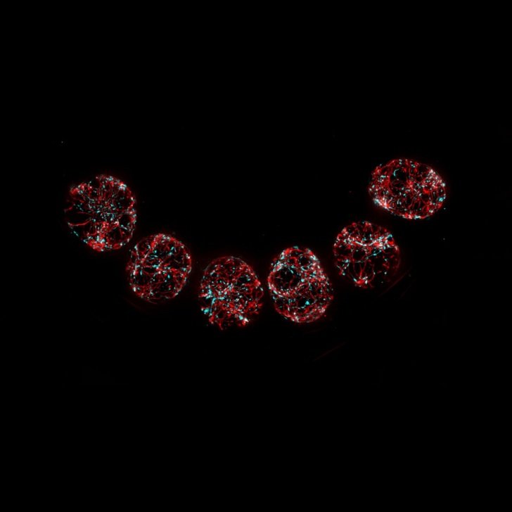
The Rivers Within
Andrew Quattrocchi, PhD student
This image shows human stem cell-derived neurovascular assembloids – miniature, laboratory grown tissues designed to mimic parts of the brain’s vascular environment. Fluorescent labels reveal branching vessel-like networks in red and supporting cells in cyan as they organise throughout each spherical assembloid. These “mini-tissues” help us study how brain cells and blood vessels develop, communicate and respond to disease.
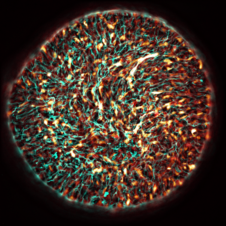
Solar Flare
Andrew Quattrocchi, PhD student
This image shows a human stem cell-derived model of the human brain. Different colours highlight the many cell types that form brain tissue and blood vessels. Their dense, intertwined networks highlight how closely brain cells and vascular cells grow together, communicate and support one another during development.
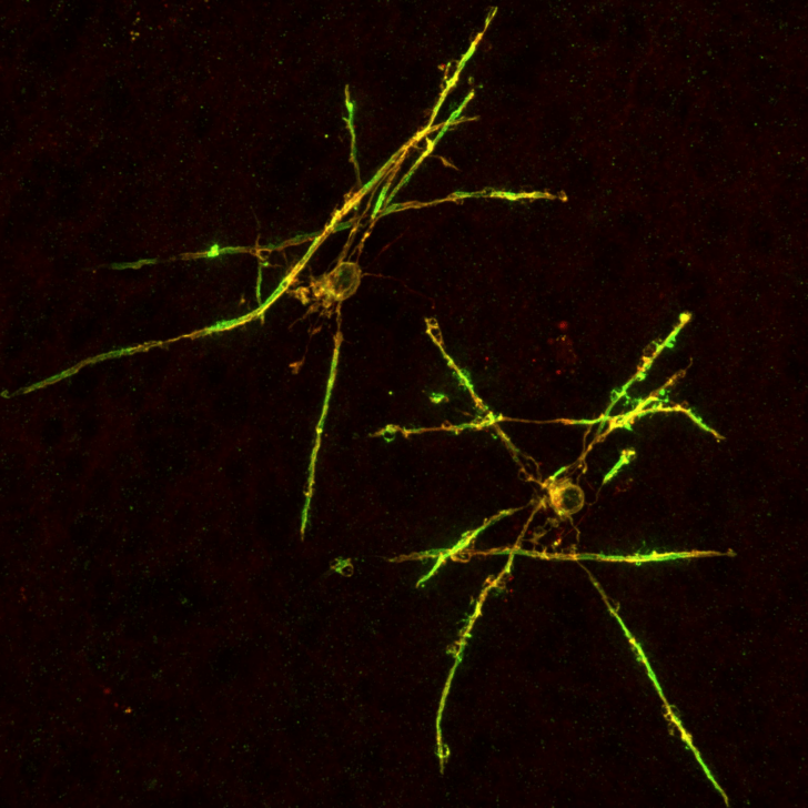
In A Gliaxy Far, Far Away...
Francois Beau, PhD student
This image shows two human oligodendrocytes and their myelin sheaths (both stained in red and green) found within a lab-grown mini human brain.
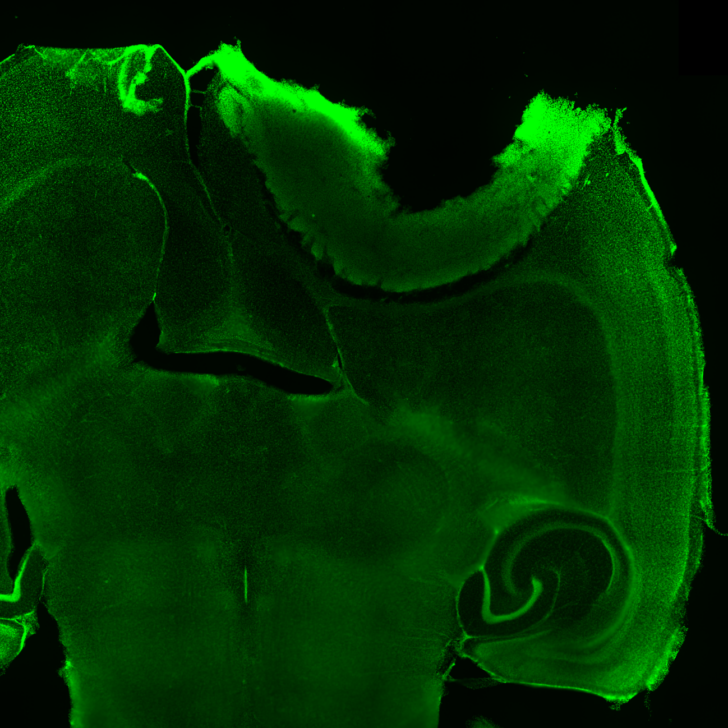
Cortex Interrupted: A Tumour's Presence in the Path of Thought
Sam Combes, PhD Student
This image shows a tumour infiltrating the mouse brain and disrupting the normal organisation of brain cells. As the tumour spreads, it can interfere with communication between neurons, which may contribute to problems with memory and other cognitive functions.
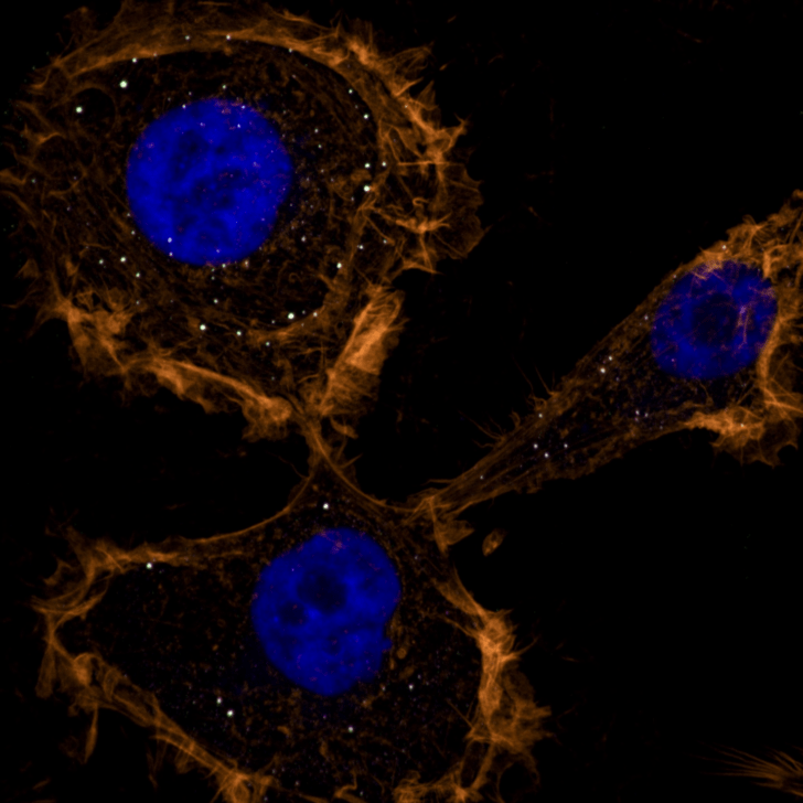
P-bodyguards
Thomas Nguyen, PhD Student
Is putting the brakes on protein production always bad? Not necessarily. These white dots are P-bodies, the cell’s emergency brakes, slowing protein production to conserve precious resources and act as bodyguards against oxidative stress.
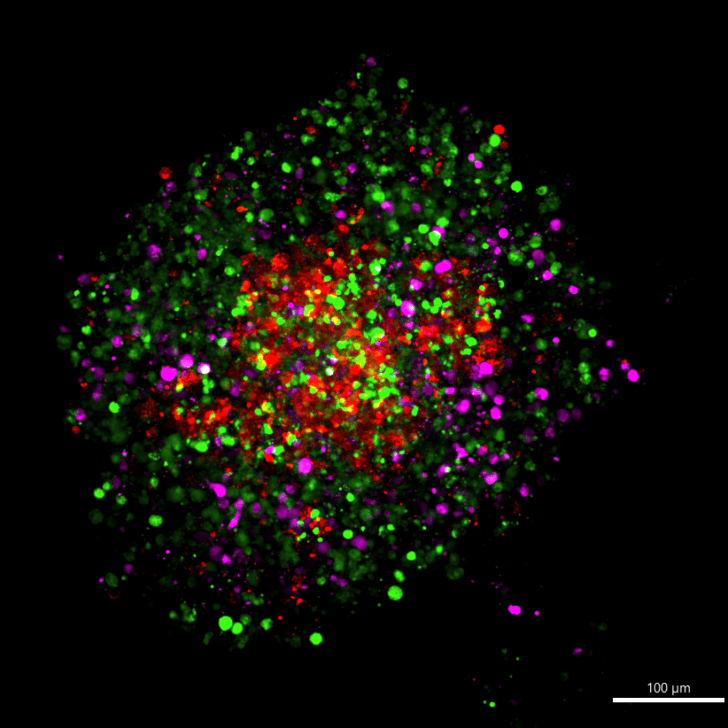
Neurovascular Galaxy
Dr Azin Amin and Dr Samaneh Mirzaei, Research Fellows
This glowing sphere is a tiny, lab-grown model of the human blood-brain barrier, the protective shield that controls what enters the brain. Magenta cells are astrocytes (support cells), green are endothelial cells (that form blood-vessel walls), and red are pericytes (that help regulate the barrier). Together they self-assemble into a 3D “neurovascular galaxy,” letting scientists study how the brain’s defences work and how diseases or drugs might affect them, all without using animal tissue.
2025 winners
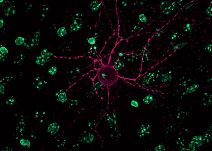
A Mind is Born
Scientific Award – James Spyrou, Neurophysiology of Excitable Networks Group
This image shows an interneuron (labelled in pink) in the cortex of the brain. All other brain cells are labelled in green.
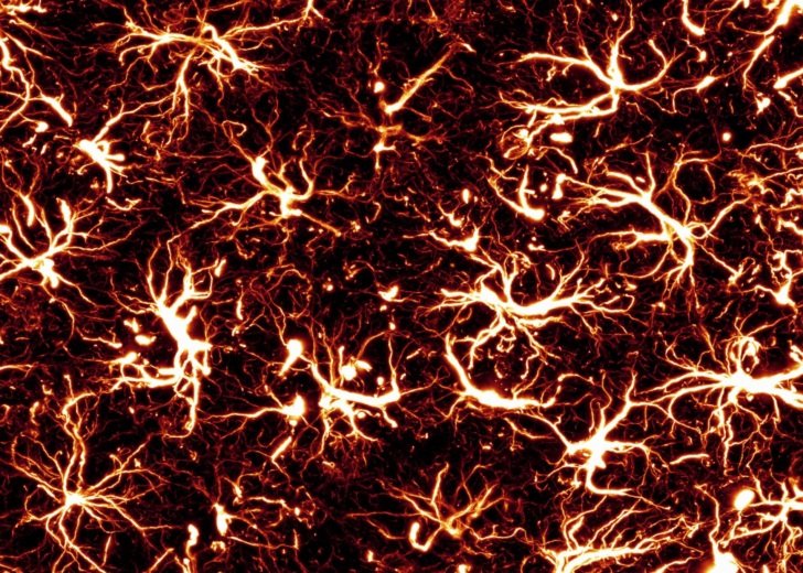
They Are Coming
People’s Choice Award – Andrew Quattrocchi, Stem Cells and Neural Repair Group
When the brain is damaged or under stress the support cells of the brain react. Here we see the chronic reaction of those support cells after a stroke.
2025 entrants
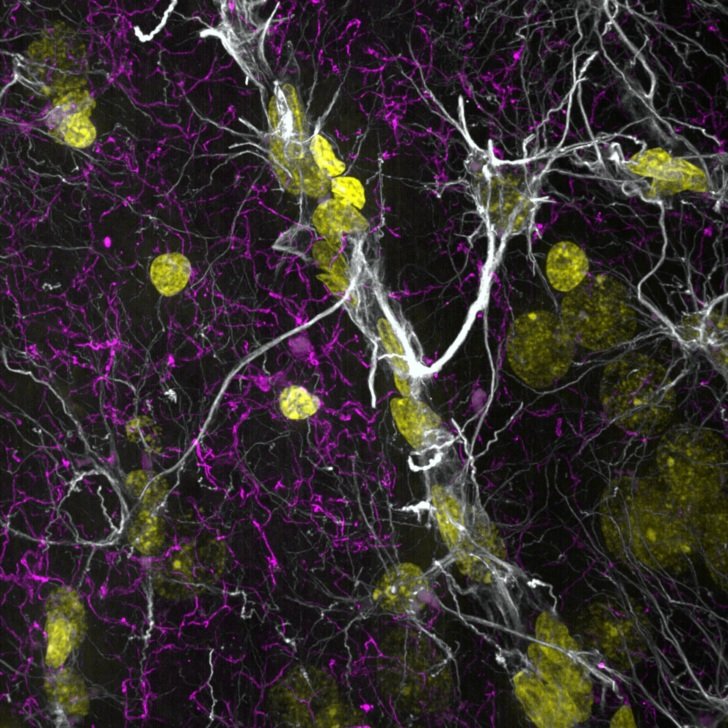
Astro Dancer
Angela Connelly, Research Assistant
This sheep forebrain image shows astrocytes in white, microglia in cyan and nuclei in yellow at 100x magnification.
Cell-fie
Chau Tran, PhD student
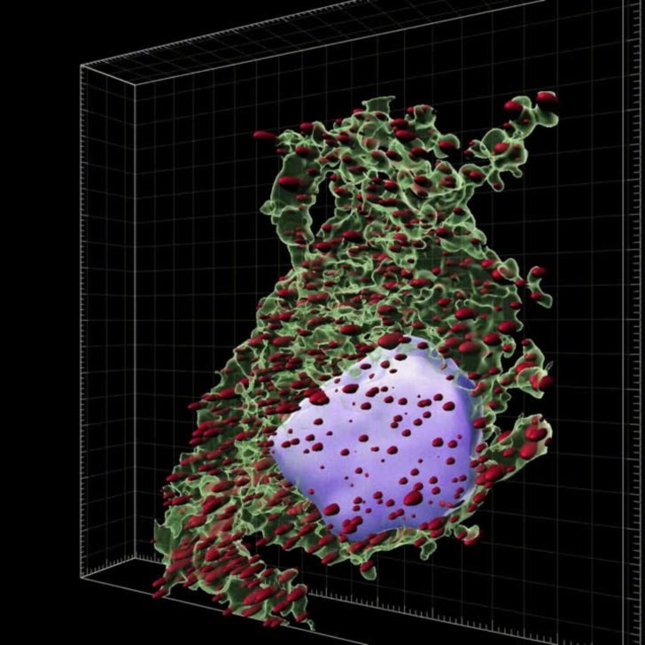
This animation shows a single human neuron that survived 9 months in a mouse brain. Despite surviving, the cell developed signs of motor neurone disease, shown as abundant puncta inside it.
Dementia Umbrella
Patricia Wongsodirdjo, PhD student
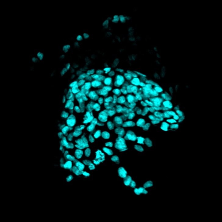
This image shows human brain cancer cells which are used in our research to test cures for dementia. The cells are labelled in bright blue.
Electric Night
Andrew Quattrocchi, PhD student
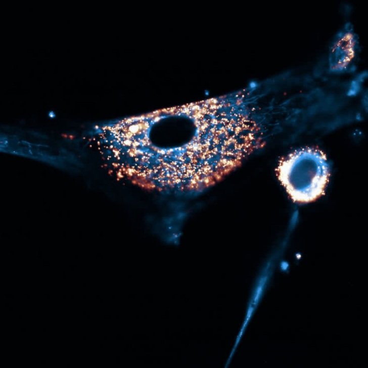
Cells have dynamic tiny machinery inside of them that allow them to function. Here we can see the mitochondria and lysosomes, the energy and recycling centers of a cell.
Hidden in Shadow
Andrew Quattrocchi, PhD student
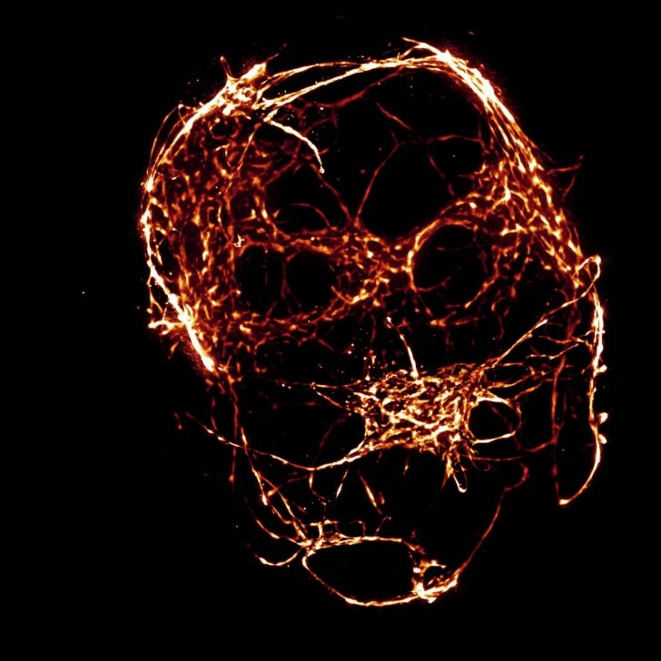
This image shows functional vessels inside a mini brain in the dish. Our ability to model the human brain is now becoming more advanced with next generation stem cell technology.
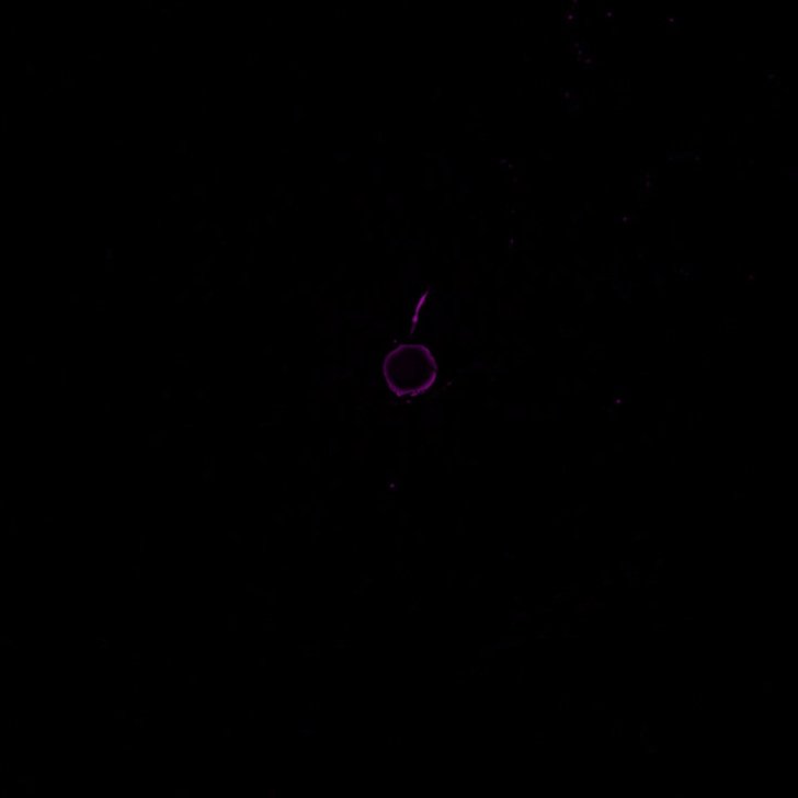
Information Superhighway
James Spyrou, PhD student
This animation shows an interneuron’s 3D structure (labelled in pink), moving up and down, illustrating what it might look like for signals to move to and from its centre.
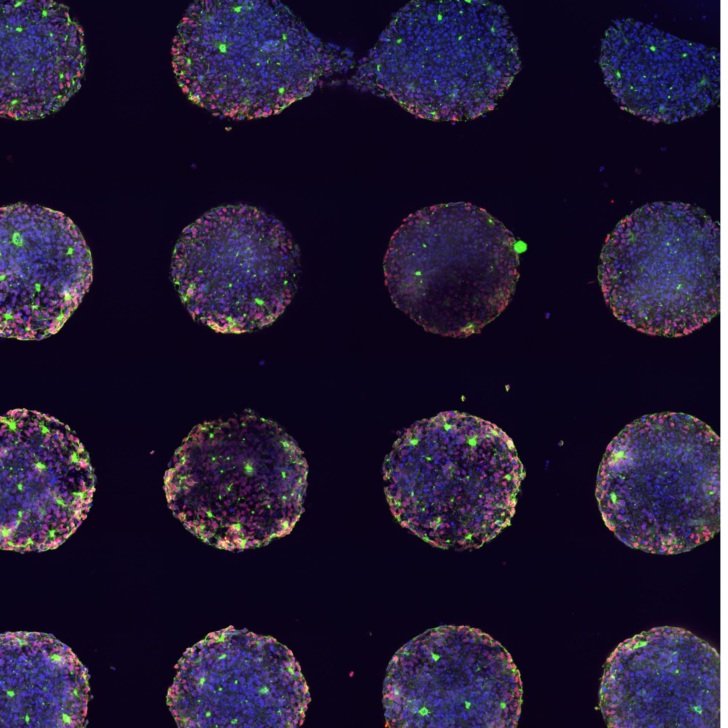
Microworlds
Montanna Waters, PhD student
This image shows stem cells that have become neural progenitor cells. The cells are grown in micro-wells, creating a uniform layout that makes them appear like planets floating in space.
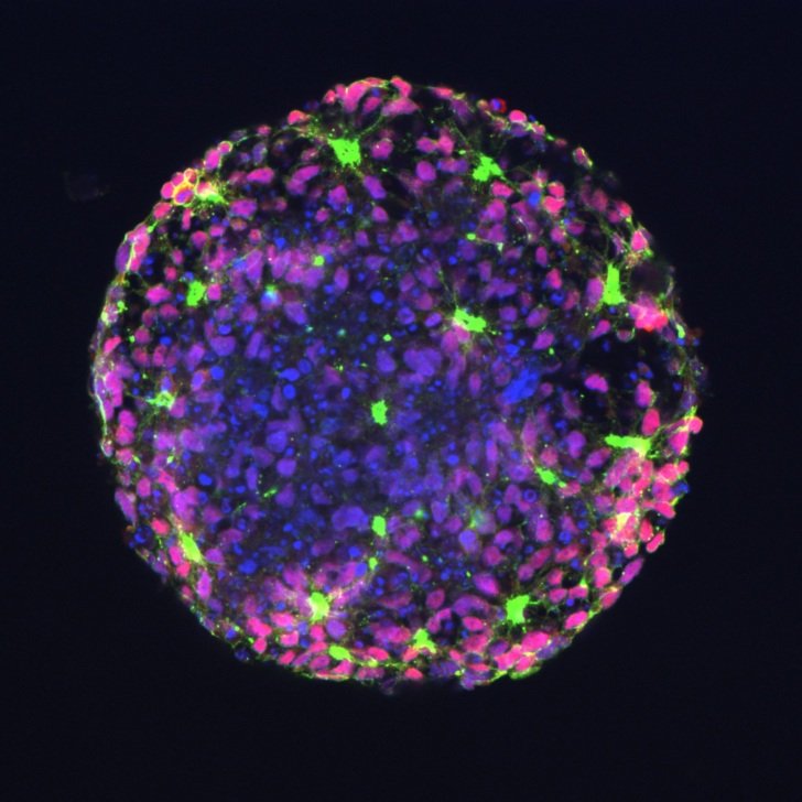
Polka-dotted Polarity
Montanna Waters, PhD student
This image shows stem cells that have become neural progenitor cells. The green staining highlights N-cadherin, a protein that helps cells stick together, the red staining indicates which cells are early brain cells, while the blue staining marks the nuclei of the cells.
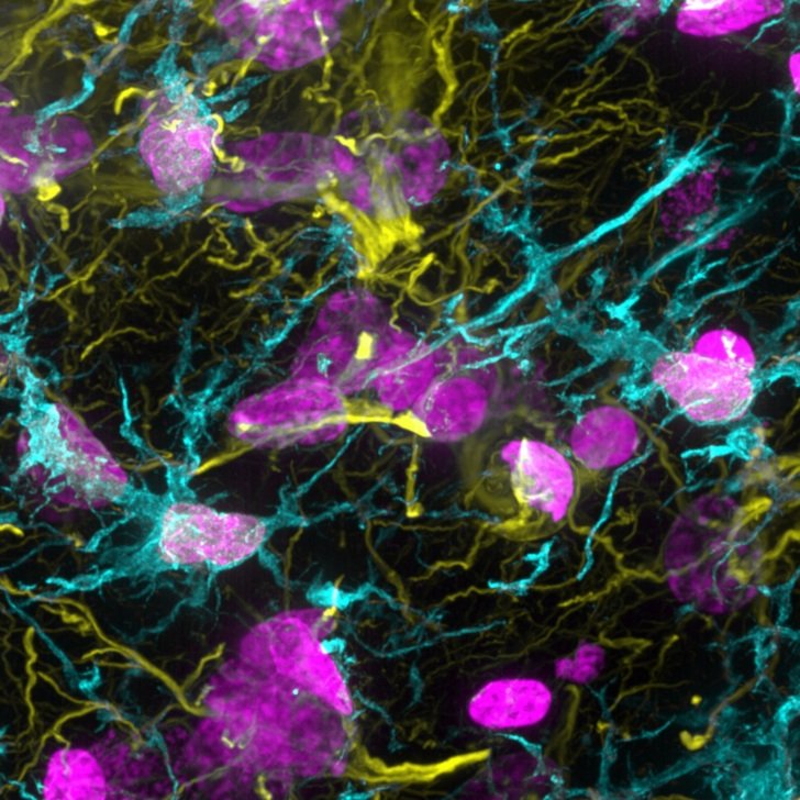
Spikeoglia
Angela Connelly, Research Assistant
This sheep forebrain image shows astrocytes in yellow, microglia in cyan and nuclei in magenta at 100x magnification.
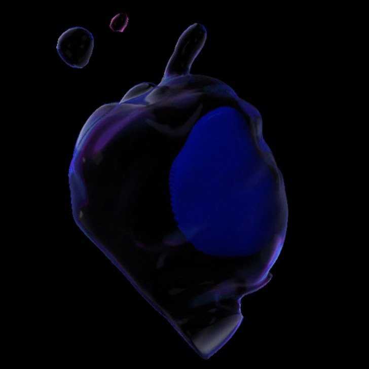
The Forbidden Cell
Chau Tran, PhD student
A 3D animation shows a human lower motor neuron. Human nuclei is labelled in blue.
2024 winners
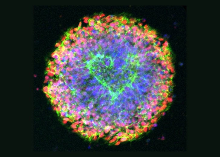
Neural Love
Scientific Award – Montanna Waters, Epilepsy Functional Genomics Group
Captured on the Nikon-Crest spinning disk confocal, this image visualises stem cells that have become neural progenitor cells. This includes early brain cells (red), cell nuclei (blue), and N-cadherin, a protein that helps cells stick together (green).
In the image, the cells have arranged around the central space in such a way where the green appears to be in the shape of a heart.
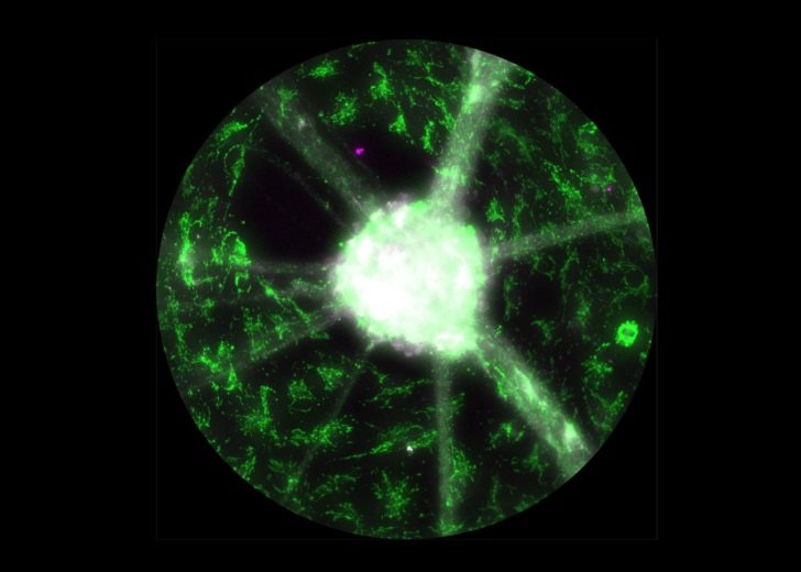
Cosmic Mitoneurons
People’s Choice Award – Chiara Parvan, Stem Cells and Neuronal Development Group
Captured on the Leica Thunder microscope, this image depicts dopaminergic neurons (neurons that undergo neurodegeneration in Parkinson’s disease) highlighted in white, mitochondria organelles within neurons in green, and pathological mitochondria in magenta.
2024 entrants
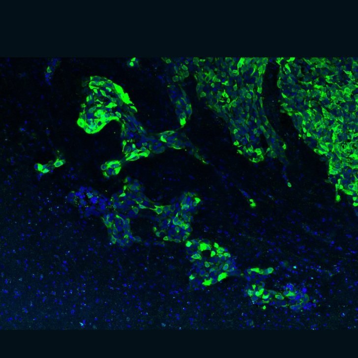
Island
Xiaoyu Wang, PhD student
Glioma cell (labelled in green) infiltrated brain. The ‘islands’ are seen on the edge of the glioma tumour mass. Brain cells are labelled in blue.
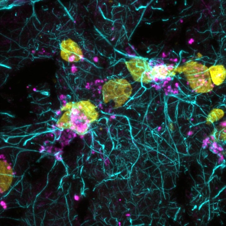
Neural Rhapsody
Chau Tran, PhD student
This image shows astrocyte (turquoise). Lipofuscin, pigment granules composed of lipid-containing residues, are labelled in pink. Cell nuclei are marked in yellow.
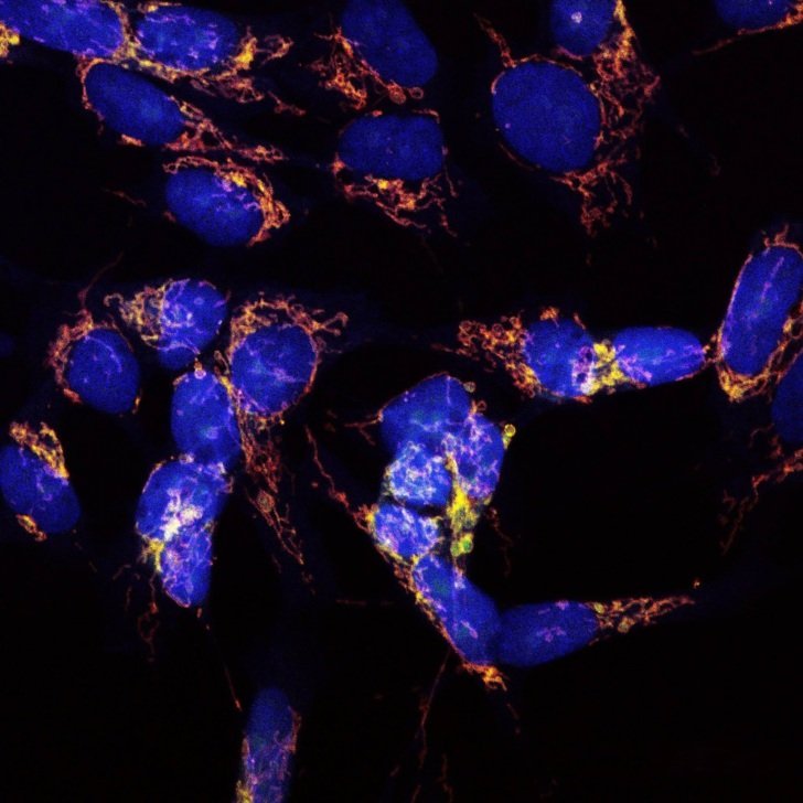
Powerhouse on Fire
Raoul Das, PhD student
Mitochondria (in red) of neurons and the immune protein Gasdermin-E (in green).
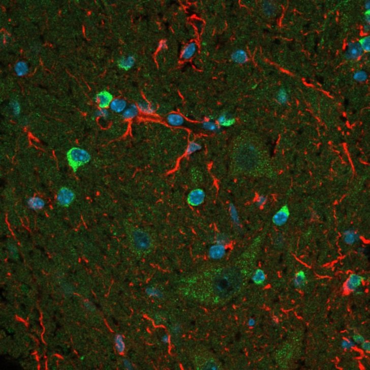
Reduce Reuse Recycle
Dushana Alagiyawanna, MPhil student
Autophagy (labelled in green by the key autophagy gene, Atg7), astrocytes (labelled in red by GFAP). Nuclei are labelled in blue.
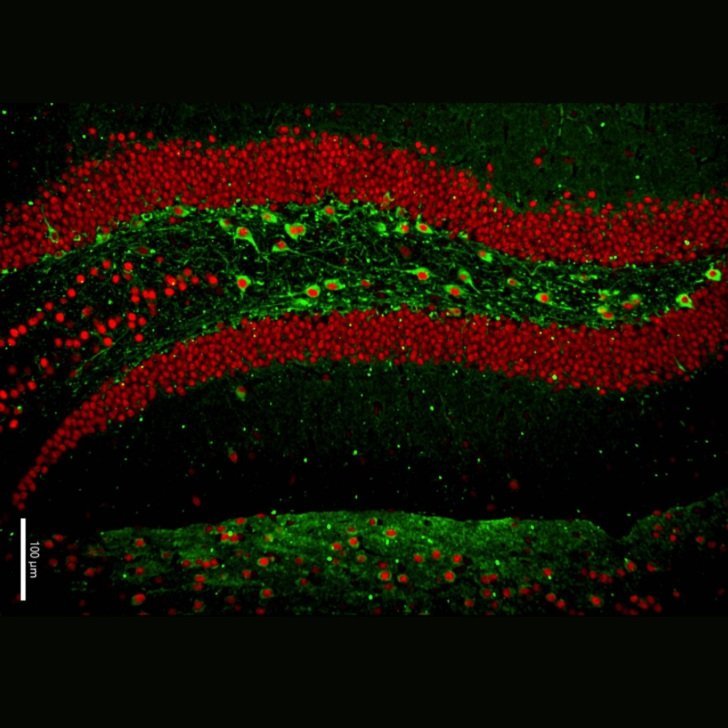
River of the Life
Samaneh Mirzaei (PhD), Research Fellow
TDP-43 proteinopathy (in red) in hippocampus and neurons in green.
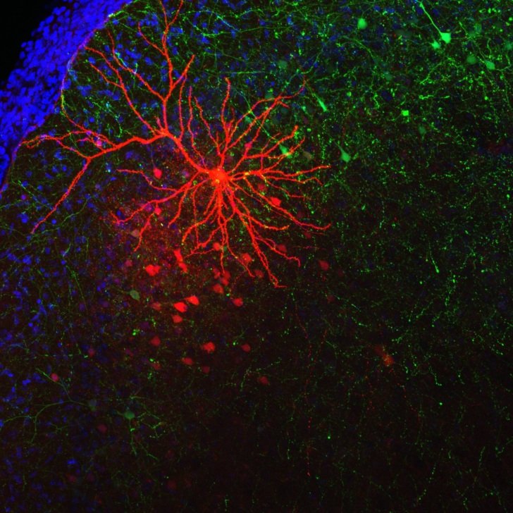
Tangled up in Red
James Spyrou, PhD student
Genetically engineered neurons (labelled in green), with the entire branches and structure of a specific neuron (labelled in red). Brain cells are labelled blue.
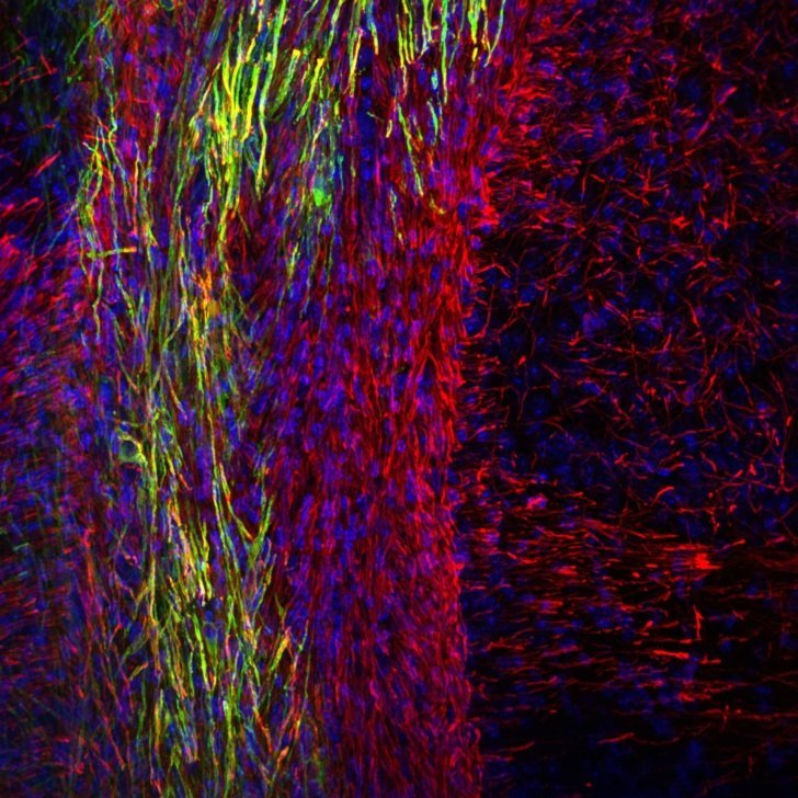
That's a Wrap!
Francois Beau, PhD student
Transplanted human myelin (green) wrapping around genetically modified brain axons.
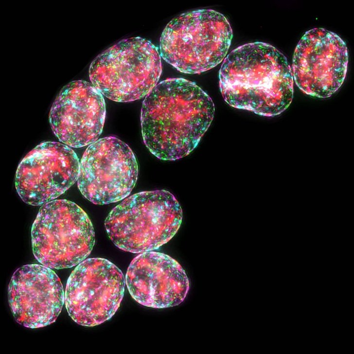
The Birth of the Brain
Andrew Quattrocchi, PhD student
Formation of brain-like systems 24 hours after formation from stem cells. The different colours show the different populations of cell types necessary for life.
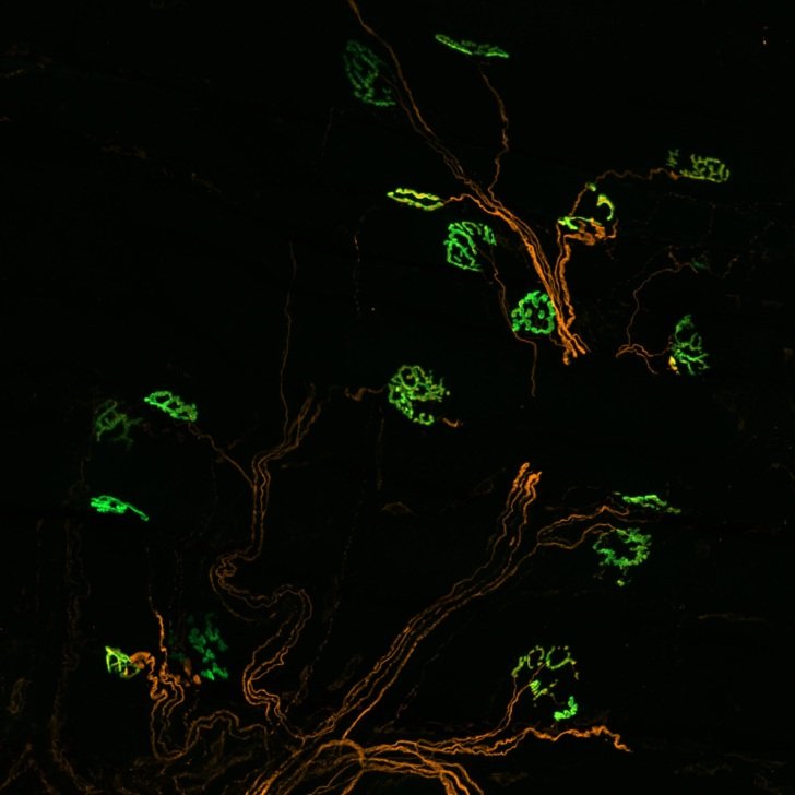
Tree of life – How Neurons Connect Us All
Aida Viden, PhD student
The bridge between nerve cells (orange) and muscle (green), the integral connections that enable us to move our muscles and bodies.
Why Aren't We Studying Morphology Instead
Lena Kricalussy-Hrabár, PhD student
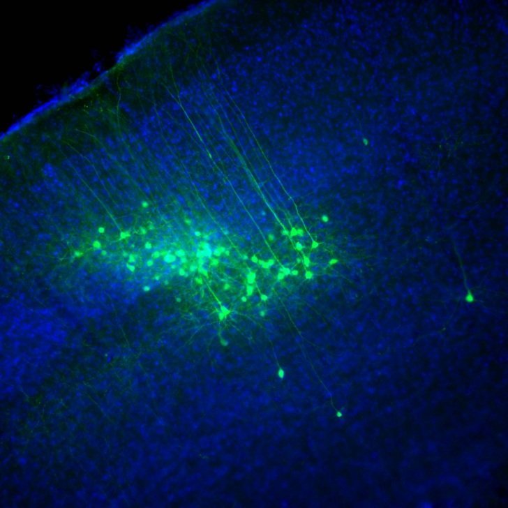
Pyramidal neurons in the cortex labelled with green fluorescent protein. These cells are one of the main cell types in our brain. All cell bodies are labelled in blue to provide a background reference.
2023 winners
Sparkling Pink Heart
Scientific Award – Shivani Vaidya, Addiction Neuroscience Group
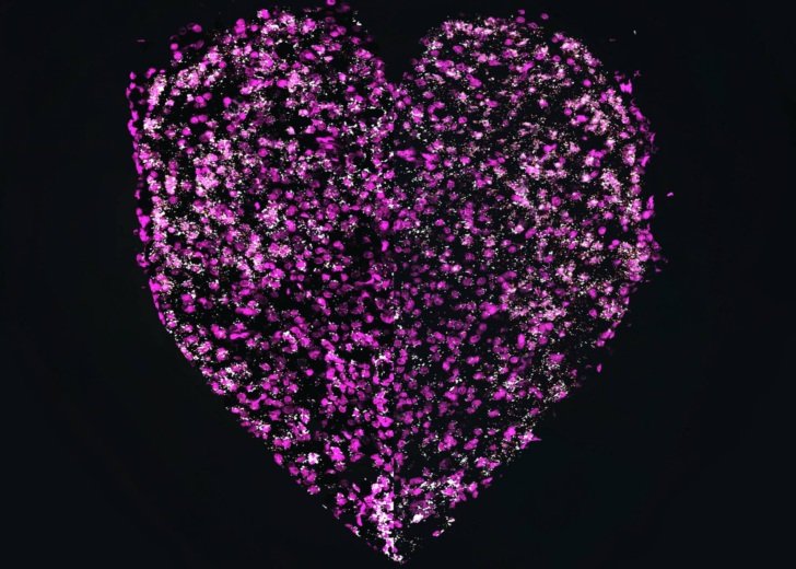
Captured on an LSM 900 confocal microscope, this combined image depicts two different hemispheres of the medial habenula.
The cell nucleus (pink) is surrounded by VGLUT1 mRNA (white).
Wysteria Joy
People’s Choice Award – Chau Tran, Personalised Therapeutics Group
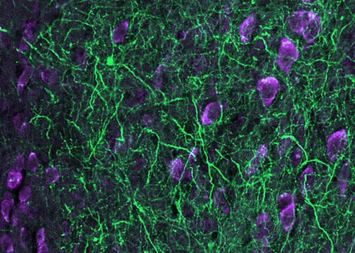
Using a Zeiss LSM 900 microscope, this image shows surviving motor neurons from patients with motor neurone disease in mouse brains after 6 months.
Lower motor neurons are labelled with CHAT (green) and purple 8-OHG, biomarker of oxidative stress.
2023 entrants
Synaptic transmission in brain organoids
Pamela Kairath (PhD), Research Fellow
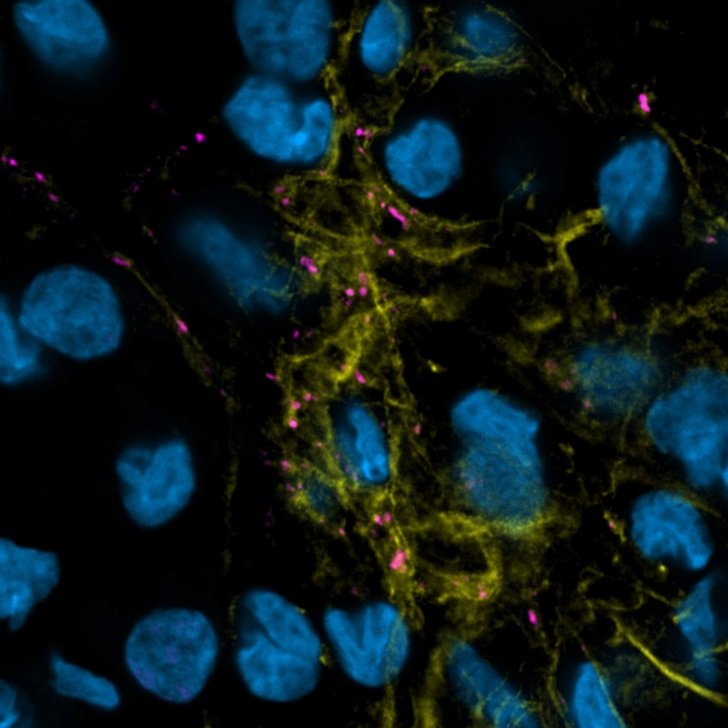
Brain organoids neurons, VGlut1, vglumate transporter 1 in pink, nuclei in blue and tubulin β3 in yellow.
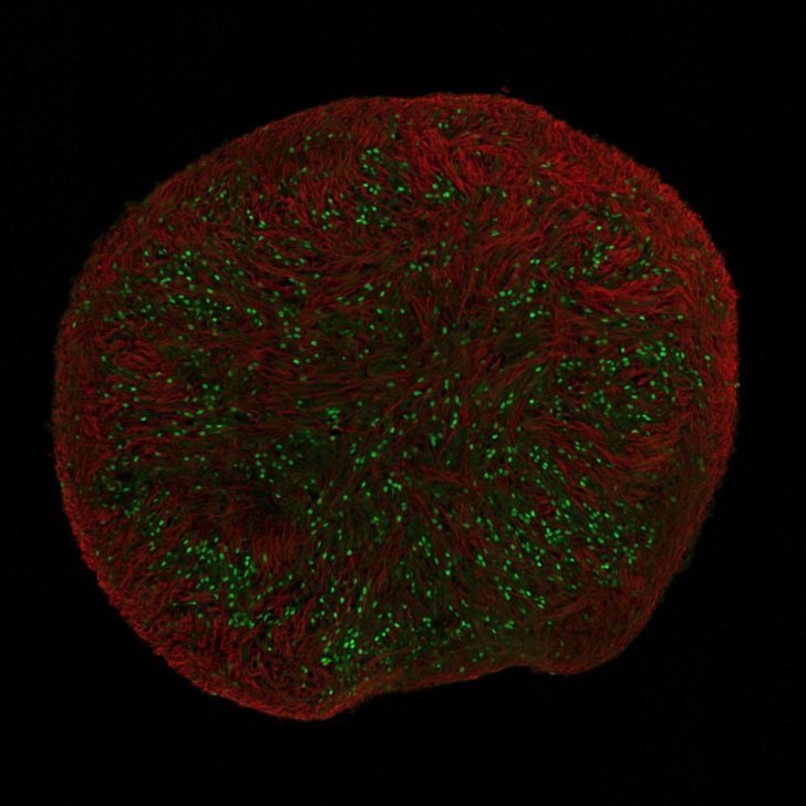
Overview of a spinal cord organoid
Lijun Loh, PhD student
Motor neurons in green and neurofilaments in red.
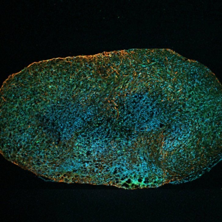
A neuromuscular organoid
Nirma Perera (PhD), Senior Research Officer
MND patient stem cell-derived organoid shows red nerve cells, blue nuclei and green autophagy protein.
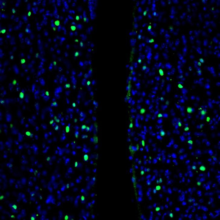
Bright cells meeting at the river of the ventricle
Alexandra Fraser, MPhil student
Stress marker c-fos in green and nuclei (blue) in a brain exposed to Porsolt test.
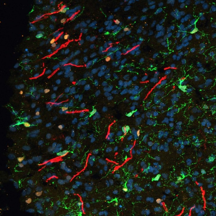
Jackson Pollock (reborn)
Francois-Xavier Beau, PhD student
Human lab-grown ‘mini-brain’. Oligodendrocytes (immature in green and mature in orange) can be seen doing their own thing (myelin in red), nuclei in blue.
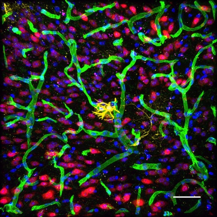
Breaking Barriers: Novel peptide-linked drugs penetrate the blood-brain barrier
Azin Amin (PhD), Research Fellow
The successful journey of drugs conjugated to a novel peptide (magenta) as they traverse the blood-brain barrier, escaping blood vessels (green) to reach the neurons (red) within the brain. Astrocytes in yellow and nuclei blue.
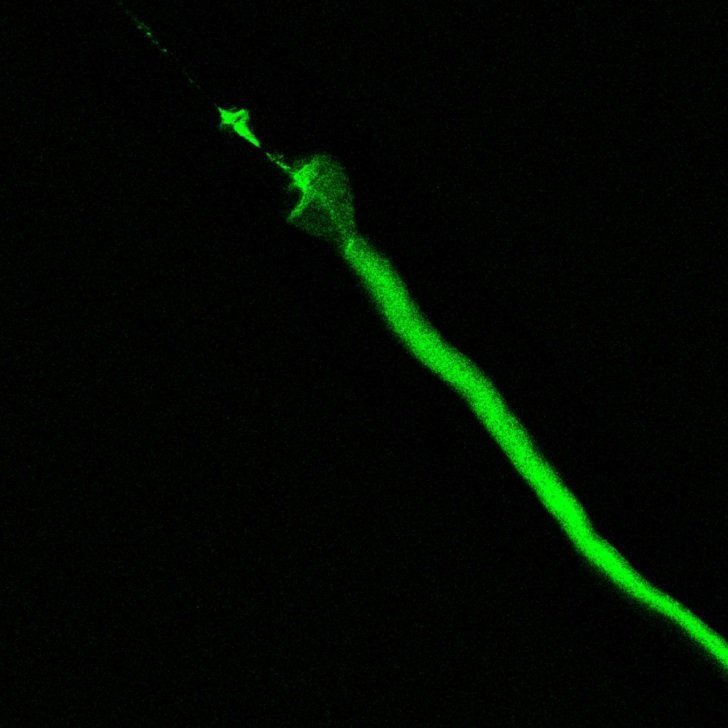
Microscopic biological sword
Hannah Truong (PhD), Postdoctoral Research Officer
Live adult C.elegans worms fed with GFP tagged Ferritin (FTN-2:GFP) and mounted onto glass slide. Head and body image was captured by SP8 confocal microscope equipped with 20x optical lens.
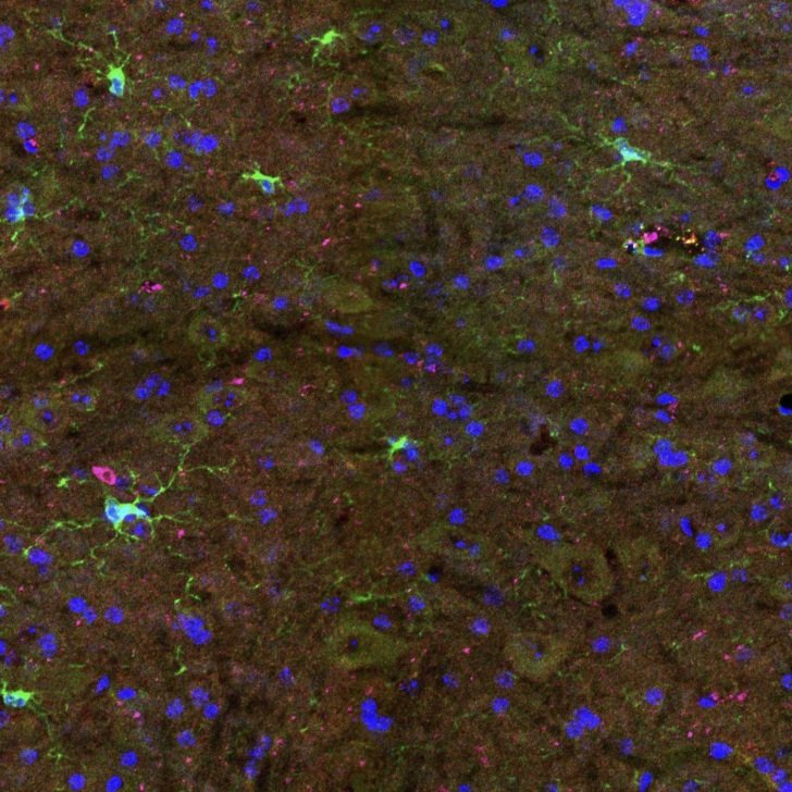
OH MY oliGODendrocyte!
Katherine (Katie) Lewis, PhD student
Oligodendroglia are the myelinating cells and are essential for maintaining brain health. Nuclei (blue), PDGFR⍺ (green; stains for oligodendrocyte precursor cells), EdU (magenta; marks cell proliferation).
Our microscopy services
The Florey Microscopy Facility supports in-house scientists and visitors in using advanced light microscopy methods for their research.
The facility houses a collection of state-of-the-art microscopy equipment and image processing tools. The experienced microscopy team are also available to assist users throughout the entire process of microscopic imaging.
All images as part of the Neuropix Imaging Competition are copyright of The Florey and cannot be reproduced without permission.