Microscope specifications
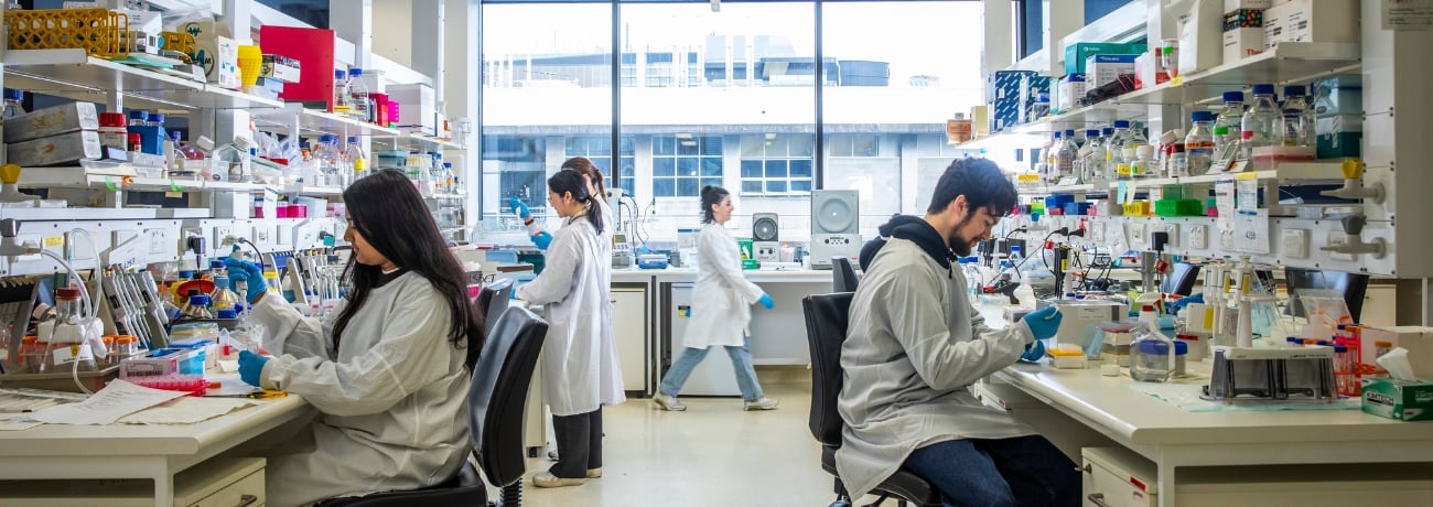
The Microscopy Facility provides advanced imaging tools and computational resources for researchers to generate detailed views of the systems and processes occurring in the brain.
Finding the right microscope for your research is paramount, the microscopy team can assist with choosing the right equipment for your needs or you can view our capabilities for the following state-of-art microscopes.
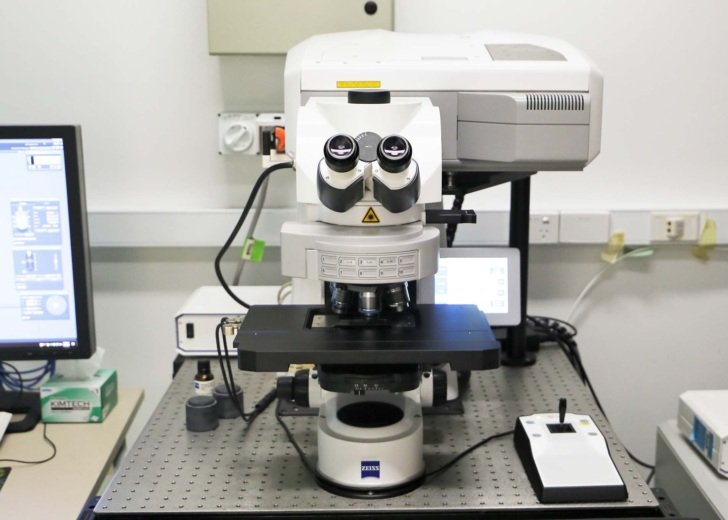
Zeiss LS M 780
Confocal laser scanning microscope used for optical sectioning, fixed tissue
The Zeiss Axio observer LSM 780 is an upright confocal microscope that can be used to image fixed cells and tissues. It is fitted with a piezo stage and can be used with a slide holder (two slides) or a universal stage insert.
A unique feature of this instrument is the simultaneous imaging of up to ten channels with the GaAsP detector array. This instrument can be fitted with long working distance 20x NA1.0 dipping lens to image cleared brain samples (can be used for SCALEVIEW as well as CLARITY cleared samples).
Applications
- z-stack
- Tile scan
- Multi positions
- Spectral unmixing
- Online finger printing
- Reflection imaging
- DIC
Light sources
- Metal halide fluorescent lamp
- Diode laser – 405 nm
- Argo laser – 458, 488 and 514 nm
- Diode pumped solid state laser – 561 nm
- HeNe laser – 594 and 633 nm
Objective details
| Objective | N.A | Immersion media | Working distance (mm) |
|---|---|---|---|
| PL-APO 10 (DIC) | 0.45 | Air | 2.000 |
| PL-APO 10 (DIC) | 0.45 | Air | 2.000 |
| PL-APO 10 (DIC) | 0.45 | Air | 2.000 |
| PL-APO 10 (DIC) | 0.45 | Air | 2.000 |
| PL-APO 10 (DIC) | 0.45 | Air | 2.000 |
Detectors
- 3 fluorescence PMTs
- 2 HyD GaAsP
- 1 transmitted light
Software
- Zen Black
Zeiss Axio Imager M2 with Apotome
The Zeiss Axio Imager is an upright microscope that can be used to image brightfield and fluorescent samples on slides. This instrument also has an ApoTome for optical sectioning of fluorescent samples.
Applications
- Brightfield imaging
- Fixed tissue imaging
- Fluorescent imaging
- Neuron tracing
- Optical sectioning with ApoTome
- Stereology
- Tile scan
- Z-stack
Light sources
- Epi-fluorescent Colibri 7.7 LED illumination
- Brightfield LED illumination
Fluorescent filter sets
| Filter cubes | Excitation | Dichromatic mirror | Emission filter |
| Filter set 90 HE LED (E) suitable for fluorescent dyes like DAPI, FITC, TRITC and Cy5 | 385, 475, 555 and 630 nm | ||
| Filter set 96 HE BFP shift free (E) | EX BP 390/40 | 420 | EM BP 450/40 |
| 38 HE eGFP shift free (E) | EX BP 470/40 | 495 | EM BP 525/50 |
| Filter set 43 HE Cy 3 shift free (E) | BP 550/25 | 570 | EM BP 605/70 |
| Filter set 50 Cy 5 shift free (E) | BP 640/30 | 660 | EM BP 690/50 |
Objective details
| Objective | NA | Immersion media | Working distance (mm) |
| EC PL-Neofluar 2.5 | 0.085 | Air | 8.80 |
| PL-APO 10 | 0.45 | Air | 2.10 |
| PL-APO 20 | 0.80 | Air | 0.55 |
| EC PL-Neofluar 40 | 1.30 | Oil | 0.21 |
| PL-APO 63 | 1.40 | Oil | 0.19 |
| PL-APO 100 | 1.40 | Oil | 0.17 |
Cameras
Lumina HR and Hamamatsu Orca-Flash 4 CMOS cameras
Stage
Motorised linear encoded XY and Z stage
Software
LuCam and HCImaging for basic image acquisition
Stereo Investigator
Neurolucida 360
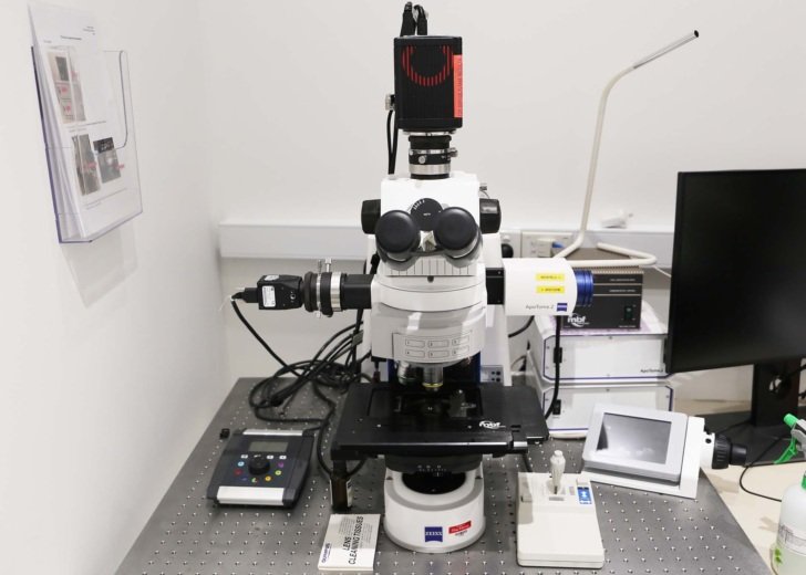
Zeiss LSM 900
Confocal laser scanning microscope used for optical sectioning and super resolution image acquisition, fixed tissue
The Zeiss Axio Observer 7 LSM 900 is an upright confocal microscope that can be used to image fixed cells and tissues. The system is fitted with a multi-immersion 25x/0.8NA objective and monochrome AxioCam 712 for fluorescent imaging too.
The unique feature of this system is the Airyscan 2, a fibre-coupled 32 array channel GaAsp detector which can capture super resolution images.
Applications
- Fixed tissue imaging
- Fluorescent imaging
- Spectral imaging
- Super resolution imaging
- Tile scan
- Transmitted light imaging
- Z-stack
Light sources
- Colibri 7
- Transmitted LED
- Main Beam Splitter (405, 488, 561, 640nm)
Filter sets
| Filter cube | Excitation (nm) | Dichromatic mirror (nm) | Emission filter (nm) |
| 90 HE LED | 382 | QBS 405 | QBP 245/30 |
| 475 | QBS 493 | QBP 514/30 | |
| 555 | QBS 575 | QBP 592/25 | |
| 630 | QBS 653 | QBP 709/100 | |
| 91 HE LED | 430 | TBS 450 | TBP 467/24 |
| 511 | TBS 538 | TBP 555/25 | |
| 590 | TBS 610 | TBP 687/145 |
Objective details
| Objective | NA | Immersion media | Working distance (mm) |
| EC PL Neofluar 5 | 0.16 | Air | 18.50 |
| PL-APO 10 | 0.45 | Air | 2.0 |
| PL-APO 20 | 0.8 | Air | 0.55 |
| PL-APO 25 | 0.8 | Water, silicone, oil, glycerol | 0.57 |
| PL APO-40 | 1.3 | Oil | 0.2 |
| PL-APO 63 | 1.4 | Oil | 0.14 |
Detectors
- Fibre-coupled 32 channel GaAsp array Airyscan 2 detector
- 2x GaAsp detectors
- ESID transmitted light detector
- Axiocam 712 mono (3.45 x 3.45µm pixels)
Software
Zen Blue
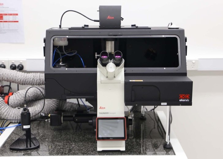
Leica Thunder
Fluorescence microscope for high throughput optical sectioning of live and fixed cells or tissue
The Leica Thunder is an inverted fluorescence microscope that can be used to image live and fixed cells and tissues. The system boasts high speed image acquisition capabilities which is useful for high throughput experiments. Interoperability with the Leica SP8 enables seamless transition of workflows from one system to another.
Features include closed loop focusing, environmental chamber, instant computational clearing and deconvolution to generate clear and crisp images.
Applications
- High throughput imaging
- Live cell imaging
- Multi positions
- Tile scan
- Time series
- Z-stack
Light source
- Epifluorescent LED 8 (395,438,475,511,555,575,635,730nm)
Fluorescent filter set
| Filter set | Excitation | Dichromatic mirror | Emission filter |
| DTF51011, size P | 391/32 | 415 | 440/40 |
| 479/33 | 500 | 510/40 | |
| 554/24 | 572 | 590/50 | |
| 638/31 | 660 | 700/75 | |
| CYR71010 | 436/28 | 459 | 473/22 |
| 506/21 | 523 | 539/24 | |
| 578/24 | 598 | 641/78 | |
| 730/40 | 763 | 810/80 | |
| 100% transmission |
Objective details
| Objective | NA | Immersion media | Working distance (mm) |
| N PL 5 | 0.12 | Dry | 14 |
| HC PL Fluotar 10 | 0.30 | Dry | 11 |
| HC PL FL L 20 | 0.40 | Dry | 7.5-6.2 |
| HC PL APO 20 | 0.80 | Dry | 0.40 |
| HC Fluotar 25 | 0.95 | Water | 2.4 |
| HC PL APO 63 (corr) | 1.4-0.60 | Oil | 0.14 |
Camera
Leica K5 sCMOS camera (2048 x 2048 resolution, 6.5 x 6.5 µm pixel)
Stage
Motorised linear encoded XY and Z stage
Software
LAS X 3D
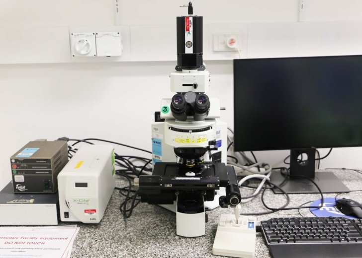
Olympus BX51
The Olympus BX51 is an upright microscope that can be used to image brightfield and fluorescent samples on slides.
Applications
- Fluorescent imaging
- Stereology
- Tile scan
- Transmitted light imaging
Light sources
- 12V/ 100W HLX Xenophot 64625 Halogen lamp (BF)
- HBO 100W Hg short arc lamp (Fluorescence)
Fluorescent filter sets
| Filter cubes | Excitation | Dichromatic mirror | Emission filter |
| UMWU2(UV) | UV wide (BP 330-385) | 400 | BA420 |
| UMNIBA2 (Blue) | Blue narrow (BP 470-490) | 505 | Bandpass (BA 510-550) |
| UMWIB2 (Blue) | Blue wide (BP 460-490) | 505 | Interference (BA510IF) |
| UMWIG2 | Green wide (BP520-550) | 565 | Interference (BA580IF) |
| U-MNG2 | Green narrow (BP530-550) | 570 | Standard BA590 |
Objective details
| Objective | NA | Immersion media | Working distance (mm) |
| PL 2 | 0.05 | Air | 5.80 |
| UPLSAPO 10 | 0.40 | Air | 3.10 |
| UPLSAPO 20 | 0.7 | Air | 0.60 |
| UPLSAPO 40 | 0.75 | Air | 0.51 |
| UPLSAPO 60 | 1.35 (DM Ph4) | Oil | 0.15 |
| UPLSAPO 100 | 1.40 | Oil | 0.20 |
Cameras
Q Imaging and Hamamatsu Orca R2 cameras
Stage
Motorised LudL XY stage
Software
QCapture and HCImaging for basic image acquisition
Stereo Investigator
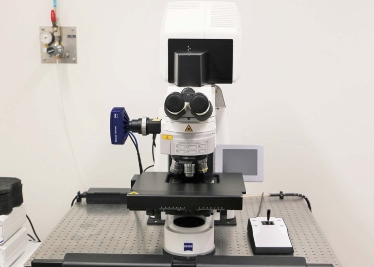
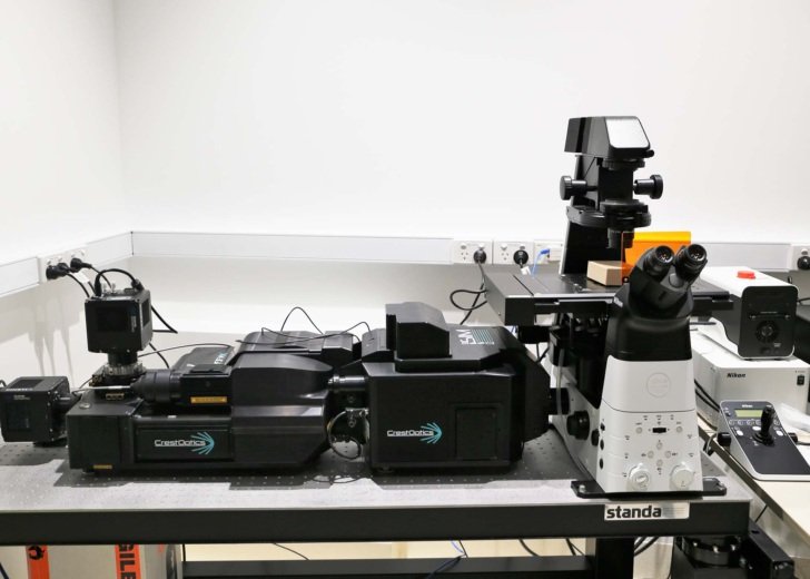
Nikon-Crest Optics Spinning Disk Confocal
An inverted microscope with Crest V3 light spinning disk and DeepSIM for both high-resolution and tissue imaging.
Simultaneous dual-channel camera acquisition with up to 25mm view.
Applications
- Confocal imaging
- Fluorescent imaging
- Imaging with large field of view
- Super resolution
- Tile scan
- Transmitted light imaging
- Z-stack
Light sources
- D-Ledi LED system (385, 475, 550, and 621nm)
- Solid state lasers 405, 488, 555, 640 and 730nm
| Filter cube | Excitation | Emission filter (nm) |
| 5-way (ZET 405/488/555/640/730 r25mm) | 405 488, 555 640 730 | 440/60 520/40 590/50 680/50 825/150 |
| ET | 440 | 40 |
| ET | 525 | 50 |
| ET | 600 | 50 |
| ET | 667 | 60 |
| ET | 811 | 80 |
| 4-way(ZET405/488/555/640) | 405 405 488 555 640 | 440/60 520/40 590/50 720/70 |
| Splitter | 565 long pass |
Objective details
| Objective | NA | Immersion media | Working distance (mm) |
| PL-APO 4 | 0.2 | Air | 20 |
| PL-APO 10 | 0.45 | Air | 4 |
| PL-APO 20 | 0.8 | Air | 8 |
| PL-APO 25 | 1.05 | Silicone | 0.55 |
| PL-APO 40 | 1.25 | Silicone | 0.55 |
| PL-APO 60 | 1.42 | Oil | 0.15 |
| PL-APO 100 | 1.45 | Oil | 0.13 |
Cameras
2 x Photometrics Kinetix CMOS (large field of view imaging, 6.5 x 6.5µm pixel)
Stage
Motorised XY and Z stage and piezo z-drive
Software
NIS Elements
Precision Analysis workstation
Image analysis workstation used for 3D analysis and quantification consisting of a PC with Imaris, FIJI, LASX Core Offline and ZEN Blue lite.
Imaris Core Facilities. Enable 3D/4D visualisation and analyses including cell counts, volume analysis and colocalisation of confocal data. Generate 3D visuals to add to presentations. Interface for customisation with Matlab, Java or Python.
FIJI is an open-source processing package used for image analysis.
Leica’s LASX Core Offline software enables manipulation and analysis of images captured on Leica systems.
Zeiss’s Zen Blue Desktop with deconvolution toolkit enables users to visualise and post-process images captured on Zeiss hardware.
PC: Windows 10, 1x Intel Xeon Gold 6246R 3.4GHz (4.1GHz Turbo, 16C), 256GB 8X32GB DDR4 3200MHz RDIMM ECC Memory, M.2 512GB PCIe NVMe Class 50 SSD (Internal PCI card), Additional storage 2 x 3.5″ 1TB 7200rpm SATA Hard Drive (configured in RAID 1 via PERC PCIe controller), NVIDIA Quadro RTX4000 8GB, iDRAC Enterprise
Offline image analysis workstation
Image analysis workstation used for 3D reconstruction, quantification and deconvolution consisting of a PC with Stereo Investigator desktop version, Neurolucida 360 lite, Huygens essential, Zen Blue and FIJI.
The desktop version of Stereo Investigator can be used to perform stereology at the PC using image stacks and virtual image slices.
- Neurolucida 360 lite is used to reconstruct neuronal structures such as dendrites and axons.
- Huygens essential can be used to post-process and deconvolve images.
- The Zen Blue lite software allows for processing and analysis of images captured on the Zeiss confocals.
- FIJI is an open-source processing package used for image analysis.
PC: 64-bit PC workstation. Intel® Quad Core Xeon Processor ES 1630 v4 3.7GHz, 10MB cache, 64GB 2400MHz. DDR4 RDIMM memory, 2 x 512GB Solid State Drives in RAID 1 configuration (total 512GB storage) and 8GB GDDR5 video card. Dual monitors.